Structural biology seeks to discover how biomolecules such as proteins and nucleic acids form complex three-dimensional structures as the basis of their function. Structure can relate to function through translating into scaffolding and 3D organization of the cell, e.g. through the dynamic organization of the cytoskeleton. Structure also enables biological macromolecules to generate biochemical microenvironments that facilitate chemical reactions. Thus, the structural organization of proteins is key to understanding our enzymatic catalysis is regulated, how individual proteins assemble into larger macromolecular complexes that perform highly specialized and regulated tasks such as transcription.
Cryo-electron microsopy has developed into a mainstream structural biology technique due to major technical advances during the last ~15 years. Not only have resolution regimes become achievable that allow conclusions at mechanistic, even near-atomic detail, but cryo-EM has expanded the availability of structural information to large, difficult to purify, and flexible complexes.
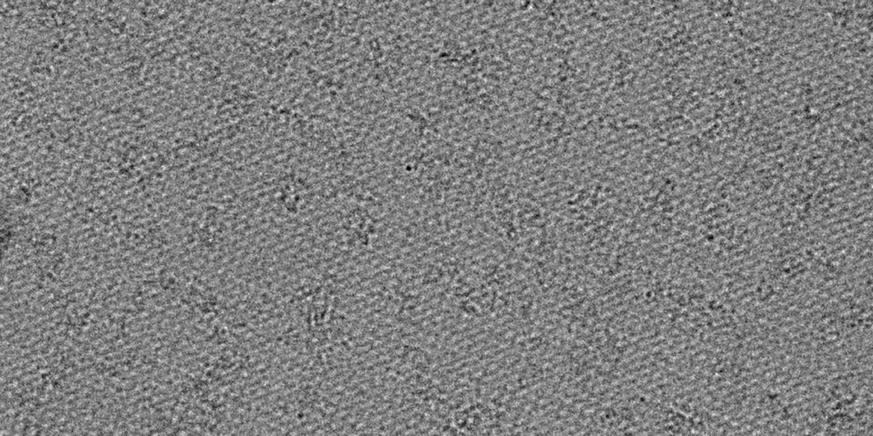
Our previous research may serve as an example of such challenging specimen. The inherent flexibility of nucleosome recognition by the human polycomb repressive complex 2 (PRC2) could only be elucidated and interpreted due to the ability of cryo-EM to visualize variability within populations of protein (-nucleic acid) complexes in solution (work in the Lab of Eva Nogales, UC Berkeley). It was therefore possible for us to explain that PRC2 is able to bind two nucleosomes simultaneosly while tolerating potential geometrical constraints imposed by the local chromatin environment. The reason for these strengths lie in the technique itself. In single particle cryo-EM, hundreds of thousands, often millions, of individual images (i.e. 2D projections) of the proteins or complexes of interest are fed into a sophisticated image processing pipeline to calculate the underlying three-dimensional shape(s) of molecules giving rise the the experimental data. Image processing tools allow researches to make use of the fact that typical datasets represent a population of molecules most often covering some degree of movements within the complex. It is possible to identify and interpret these flexibilities that can reflect important aspect of the function or regulation of the molecules of interest.
At the CMMC and on the Campus of the Medical and Natural Sciences Faculties of the University of Cologne, the infrastructure for high-resolution single particle cryo-EM is available or in the build-up. The actual data collection and processing is one of several work steps in the effort towards elucidating the structure and function of biomolecules. In our lab, we strive to cover all steps, including protein expression, purification, activity assays, cryo-EM sample preparation, screening, data collection, processing and interpretation, as well as, where possible, cellular and genomic assays. Thereby, we aim to gain a comprehensive understanding of our biological systems and provide a rich learning opportunity for students and experienced scientists who join our laboratory. We find that this ambitious aim justifies the extra effort required to establish all the relevant methods in our group. Fortunately, the CMMC has commited to supporting this endeavour and enables productivity through providing the equipment and access that we need. For example, the Tergeo plasma cleaner allows to treat even the most sensitive samples with highly controlled plasma, and using the Mark IV Vitrobot by Thermo Fisher, we can vitrify samples in our laboratory. Sample screening is possible at the CECAD imaging facility, where we have access to a 200 kV JEOL JEM2100PLUS, cryo compatible microscope that is equipped with a Gatan OneView CCD camera and SerialEM for automated data collection (https://www.cecad.uni-koeln.de/research/core-facilities/imaging-facility/). Moreover, a cryo-EM facility that is going to be opened in 2020 provides access to a state-of-the-art 300 kV Titan Krios microscope for large scale, high resolution data collection. Data processing is enabled through a high performance GPU workstation in house, as well as access to GPU compute nodes housed in the HPC Cluster at the University of Cologne (https://rrzk.uni-koeln.de/) as well as their CPU infrastructure.